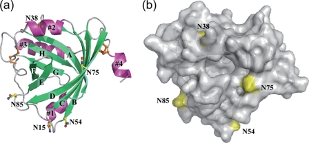
FIGURE 1.
a, crystal structure of the A variant (C149R) of hAGP at a resolution of 2.10 Å shown as a ribbon diagram. Secondary structures are colored lime green (β-strands) and light magenta (α-helices). Cys residues are depicted as orange sticks and balls. Asn residues of five N-linked glycosylation sites are depicted as yellow sticks and balls. Nitrogen and oxygen are colored blue and red, respectively. b, top view into the β-barrel with surface representation. Asn residues corresponding to glycosylation sites are colored yellow.

