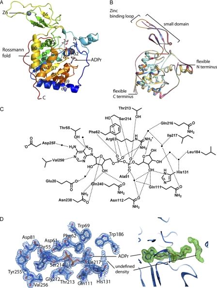
FIGURE 2.
Structure of human SIRT6 in complex with ADP-ribose. A, overall structural features of SIRT6 monomer. B, superimposition of the six molecules in the asymmetric unit. Red, chain A; green, chain B; dark blue, chain C; orange, chain D; cyan, chain E; yellow, chain F. C, schematic illustration of the hydrogen bonding network surrounding ADPr; hydrogen bonds are indicated as dashed lines, and water molecules are shown as spheres. D, left, SIRT6·ADPr 2Fo − Fc electron density map (blue mesh, 1.5σ) of the residues within 4 Å of ADPr. Right, Fo − Fc omit electron density map (green mesh, 2σ) of the ADPr molecule; the putative peptide binding site contains an unidentified electron density.

