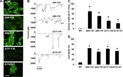
FIGURE 6.
Deletion of charged or hydrophobic residues in the central region causes toxicity. A, shown is surface immunofluorescence staining of PrP on HEK cells stably expressing Δ94–134, Δ94–110, Δ111–134, or Δ114–122 PrP. Scale bar = 50 μm. B, whole-cell patch clamp recordings at −80 mV were made from cells expressing the indicated constructs. C, quantitation of the currents recorded in panel B are plotted as the percentage of total time the cells exhibited an inward current ≥450 pA (mean ± S.E., n = 5 cells). D, cell death induced by G418 treatment (400 μg/ml) was measured by MTT reduction (mean ± S.E., n ≥ 10 wells from 3 independent experiments). Asterisks (*) indicate values that are significantly greater than those for WT PrP (p < 0.05, one-tailed Student's t test).

