
Figure 10.
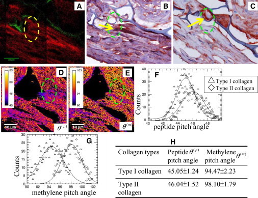
Localizations of type I and II collagens in engineered cartilage tissues through immunohistochemical staining, peptide, and methylene pitch-angle imaging. (A, Red) SHG from collagen and (A, green) autofluorescence from chitosan scaffold. (B and C) Immunohistochemical images for type I and II collagen, respectively. (Yellow arrows) Type I collagen (B) and type II collagen (C). (D and E) Peptide and methylene pitch-angle imaging. (F and G, histograms) Peptide and methylene pitch-angles in the dashed-circled region, respectively. (H) Summary of the peptide and methylene pitch-angles for type I and II collagen in engineered cartilage tissue.