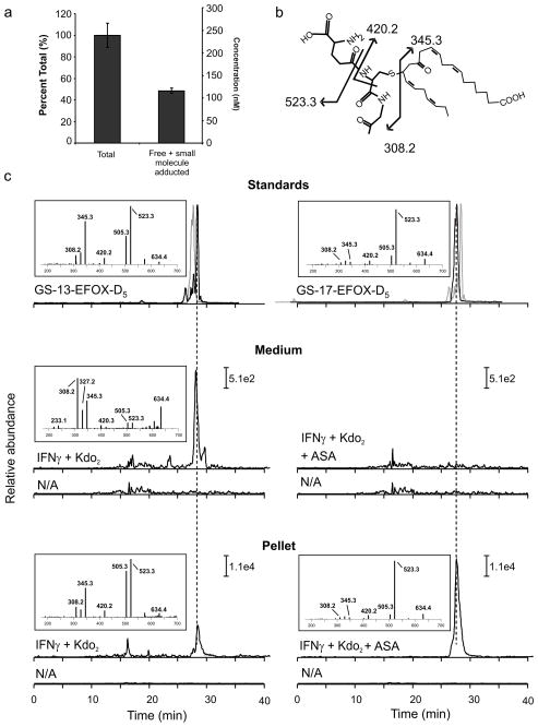
Figure 4. EFOXs form adducts with proteins and GSH following activation of RAW264.7.
a, Cell lysates from activated RAW264.7 cells were split into two groups: treatment with BME followed by protein precipitation with acetonitrile (“Total”) and protein precipitation followed by BME treatment (“Free + small molecule adducted”). b, Chemical structure and fragmentation pattern of GS-13-EFOX-D5. c, Chromatographic profiles and positive ion mass spectra of GSH adducts of 13-EFOX-D5 and 17-EFOX-D5 derived from standards (upper panels), cell medium (middle panel) and cell pellet (lower panel), N/A profiles correspond to non-activated cell samples. Grey chromatograms represent GS-17-EFOX-D5 (upper panel left) and GS-13-EFOX-D5 (upper panel right) standards. Fragments 345.3 and 523.3 were monitored in cell media and cell pellet samples respectively.

