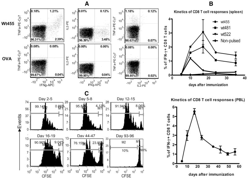
FIGURE 2.
A, CD8 T cells activated by muTRP1-lv immunization can produce multiple cytokines. CD8 T cells from muTRP1-lv-immunized mice were restimulated ex vivo with TRP1 wt455 peptides or control OVA SIINFEKL peptides and then stained for IFN-γ, TNF-α, and IL-2. Cells shown were gated on CD8 T cells. B, TRP1-specific CD8 T cell response induced by lentivector immunization is long-lasting. C57BL/6 mice were immunized with 2.5 × 10⁷ TU of lentivector muTRP1-lv. At different time points, peripheral blood cells were stimulated with pooled peptides of 455, 481, and 522, whereas the splenocytes were stimulated with each individual wtTRP1 peptides for 3 h ex vivo. Cells were then intracellularly stained for IFN-γ. The percentages of IFN-γ-producing CD8 T cells of total CD8 T cells were calculated and are presented as means plus SE. This experiment was repeated twice. C, Ag presentation in vivo after lentivector immunization is protracted. C57BL/6 mice were immunized with 2.5 × 10⁶ TU of lentivector OVA-lv. At indicated time points, mice were injected with purified CFSE-labeled OT-I cells. Three days after OT-I injection, OT-I T cell proliferation in the draining lymph nodes was analyzed by progressive banding of CFSE intensity. Numbers in each figure indicated the percentage of nondividing injected CFSE+ cells (right) and dividing CFSE-diluted cells (left). The in vivo Ag presentation assay was repeated twice.

