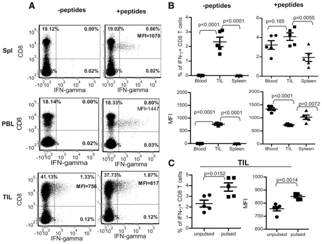
FIGURE 6.
TILs are functional and capable of producing IFN-γ after ex vivo stimulation. TILs in the tumor single-cell suspension was prepared as those in Fig. 5 and stimulated ex vivo for 3 h with tumor cells or together with additional exogenous TRP1 peptides. Additionally, PBLs and splenocytes were also collected and stimulated for staining of IFN-γ. A, Representative flow cytometry data of five mice. For analysis of TILs, gates were set in the forward scatter and side scatter dot plots by using splenocytes. Thus, most tumor cells were gated out in the TIL samples. B, Summary of the comparison of IFN-γ production among CD8 T cells from spleen, peripheral blood, and TILs, which were pulsed with and without additional exogenous TRP1 peptides. Percentages of IFN-γ+ CD8 T cells (top) and MFI of the IFN-γ (bottom) are shown. C, Summary of the IFN-γ production of TIL with or without exogenous wtTRP1 peptides. Percentages of IFN-γ+ CD8 T cells (left) and MFI of the IFN-γ (right) are shown. This experiment was repeated three times with similar results.

