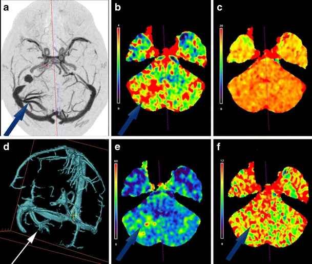
Fig. 4.

(a) A 64-year-old male with a subcortical right cerebellar hemisphere DVA on the right side (case 3) on axial DSA. Increased CTP values are present in the affected hemisphere (arrows) for regional CBV (b), regional CBF (e), and MTT (f) relative to the unaffected hemisphere. No significant differential TTP values were present (c). Orthogonally edited CTA-V view of the venous system, demonstrating the cerebellar hemisphere DVA on the right side with dilated intramedullary veins that converge into a collecting vein that enters the lateral aspect of the right transverse venous sinus is demonstrated (d)
