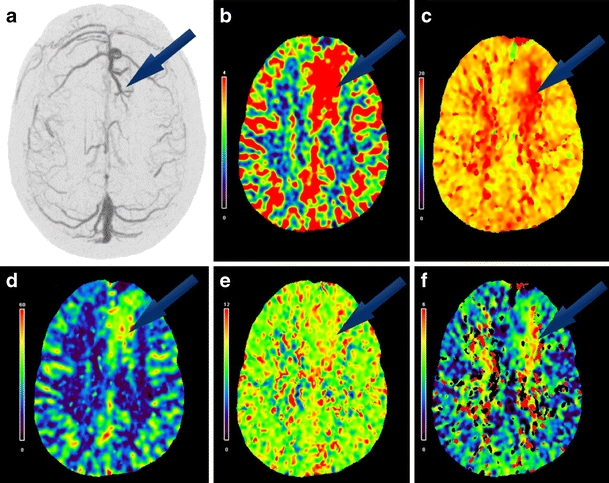
Fig. 5.

A 50-year-old female who presented with numbness on the right side of her face (case 1); a juxtacortical left frontal lobe DVA was identified. a Upper left axial image is a DSA image collected with slice thickness of 30 mm and then clockwise from left to right axial perfusion images are colorimetric representations of b regional CBV, c TTP, d regional CBF, e MTT, and f delay
