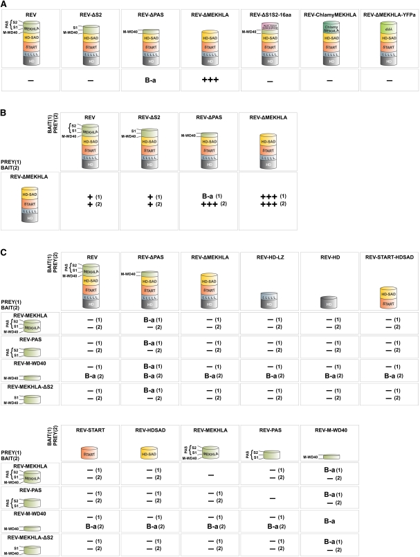
Figure 3.
The REV MEKHLA Domain Sterically Inhibits REV Homodimerization in Yeast.
Strong, weak, and no interactions are indicated by three plus signs, one plus sign, and a minus sign, respectively. Interaction strengths were determined by comparison to a set of standards (see Supplemental Figure 5 online). Reciprocal bait/prey couples are indicated by a number in parenthesis. B-a, bait autoactivation.
(A) Homodimerization Y2H assays. The same proteins were used as bait (fused to GAL4 DNA binding domain) and prey (fused to GAL4 activation domain).
(B) Interaction of REV-ΔMEKHLA with REV truncated for subsections of the MEKHLA domain.
(C) Interaction of isolated MEKHLA variants with REV protein domains.
[See online article for color version of this figure.]

