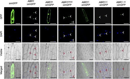
Figure 4.
Subcellular Localization of ABR1.
GFP fusions of full- or partial-length ABR1 were transiently transformed into onion epidermal cells. The overall schematic structures of each construct are shown in Figure 3A with the addition of a GFP fusion motif at the 3′ termini. The plant nuclei were stained with DAPI. Images were taken using confocal microscopy (GFP fluorescence, green; DAPI fluorescence, blue; visible, visible light image; merged, merged images of above three images). Empty vector (smGFP) transformed cells are shown as a control. Arrows indicate ABR1-localized nuclei. Bar = 100 μm.
[See online article for color version of this figure.]

