Figure 5.
DUO1 Binds in Vitro to a MYB Binding Site in the MGH3 Promoter.
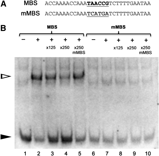
(A) Sequences of the oligos used in the EMSA experiments. MBS is the region flanking MYB site A in the MGH3 promoter (bold and underlined), and mMBS is a mutagenized version in which the MYB site has been ablated (underlined).
(B) EMSA experiments using recombinant DUO1 MYB domain with MBS or mMBS oligos. – and + indicate the absence or presence of the DUO1 MYB domain, respectively, and ×125, ×250, and ×250 indicate competing unlabeled oligos. Addition of DUO1 MYB protein (lane 2 to 5) causes a clear shift (white triangle) accompanied by a reduction in free probes (black triangle), with free probes increasing in the presence of more competing MBS oligos. In lanes 7 to 10, no shift was observed in EMSA experiments using mMBS oligos. Results were confirmed in independent EMSA experiments.

