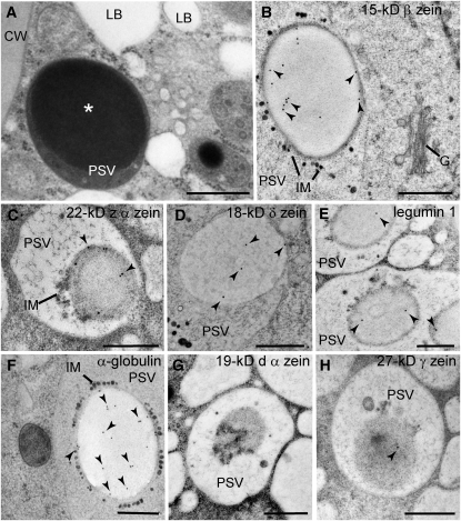
Figure 2.
Localization of Storage Proteins in Aleurone PSVs.
(A) Electron micrograph of an aleurone cell at 22 DAP containing lipid bodies (LB) and PSVs with large inclusions (asterisks). CW, cell wall.
(B) to (H) Immunogold labeling of aleurone cells at 22 DAP ([B], [D], and [F]) and 18 DAP ([C], [E], [G], and [H]) with antibodies against maize storage proteins. Most of the signal was detected on PSV inclusions (arrowheads). G, Golgi; IM, intravacuolar membranes.
Bars = 500 nm.

