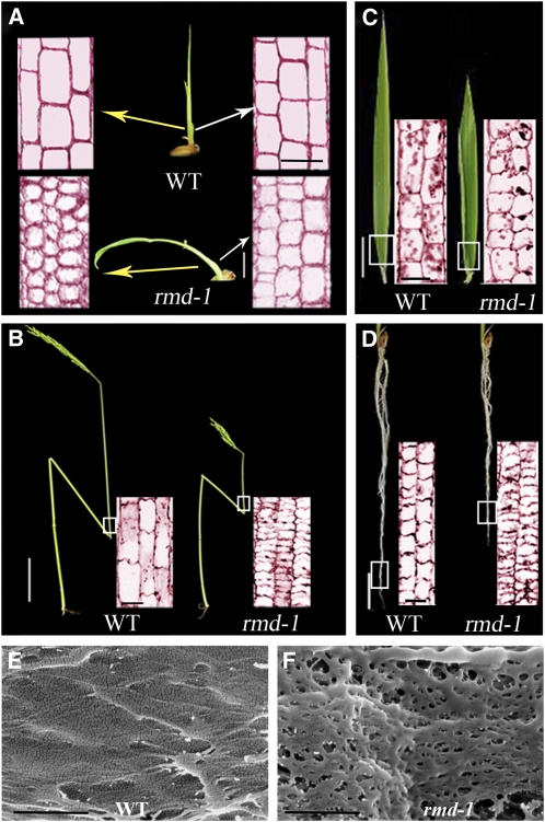
Figure 2.
rmd-1 Mutants Display Defects in Cell Elongation and Polarity as Revealed by Longitudinal Section Analysis.
(A) Leaf sheath from 12-d-old wild-type (WT) and rmd-1 seedlings. Seedling bar = 8 mm; sectioning bar = 50 μm.
(B) Culm from plants at the heading stage. Stem bar = 8 cm; sectioning bar = 100 μm.
(C) Leaf from plants at the heading stage. Leaf bar = 1.6 cm; sectioning bar = 100 μm.
(D) Roots from 14-d-old seedlings. Root bar = 5 mm; sectioning bar = 100 μm.
(E) and (F) Field emission scanning electron microscopy of the cell wall surface from epidermal cells in the root elongation zone from 14-d-old wild-type (E) and rmd-1 seedlings (F). Bars = 500 nm.
[See online article for color version of this figure.]

