Figure 3.
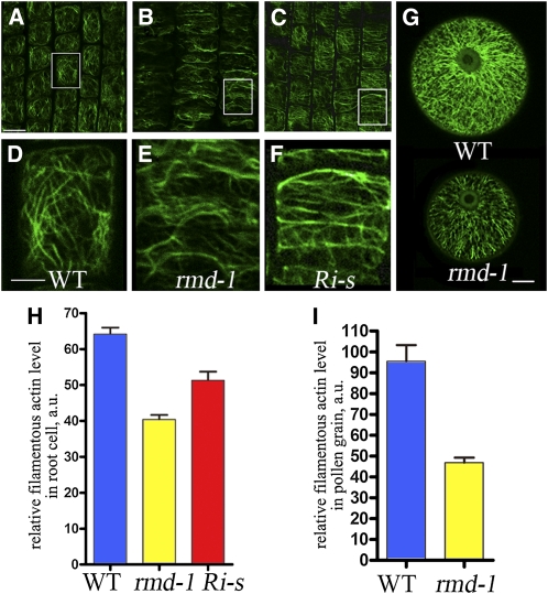
rmd Mutants and FH5/RMD RNAi Lines Display Defective Actin Microfilament Networks.
(A) to (F) Actin filaments of cells from the root elongation zone of 7-d- old rice seedlings, stained by Alexa Fluor 488-phalloidin.
(A) Wild-type cells.
(B) rmd-1 cells.
(C) Cells of FH5/RMD RNAi lines with strong defects (Ri-s).
(D) A close-up of boxed region of (A). WT, wild type.
(E) A close-up of boxed region of (B).
(F) A close-up of boxed region of (C).
(G) F-actin pattern in pollen grains of the wild type and rmd-1 at stage 13 of anther development.
(H) Fluorescent pixel intensities (±se) of wild-type, rmd-1, and Ri-s root cells were 64.16 ± 3.72 (n = 32), 40.42 ± 2.46 (n = 30), and 51.3 ± 5.05 (n = 20).
(I) The average fluorescence pixel intensity (±se) in each pollen grain was 95.48 ± 7.76 (n = 18) in the wild type and 46.87 ± 2.36 (n = 17) in rmd-1 (P < 0.001).
Bars = 50 μm in (A) to (C), 25 μm in (D) to (F), and 10 μm in (G).
[See online article for color version of this figure.]

