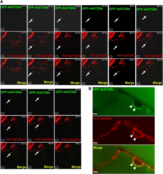
Figure 9.
YFP-AtTSPO Partially Colocalizes with GFP-AtATG8e in Plant Cells.
(A) Confocal images of a time-lapse study on root cells of a tobacco plant stably coexpressing YFP-AtTSPO and the autophagy marker GFP-AtATG8e. The arrow indicates a moving autophagosome (GFP-AtATG8e fluorescence, green channel, and merged) containing YFP-AtTSPO fluorescence (red channel and merged) seen from time point 22.1 to 36.9 s. Bar = 5 μm.
(B) Similar sample to that in (A) but imaged after concanamycin A treatment of the root cells. Diffuse GFP-AtATG8e fluorescence is visible in the vacuole (green channel), and brighter areas are seen around the nucleus (arrowheads) colocalizing with YFP-AtTSPO (red channel and merged image). Bar = 5 μm.

