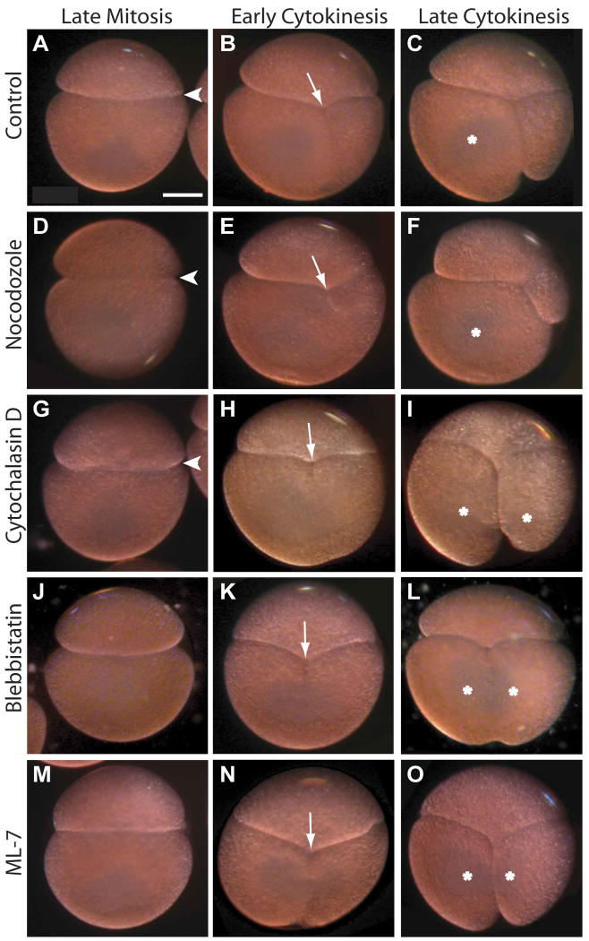
Figure 6. Actomyosin inhibitors block asymmetrization of the AB/CD interface.
Representative images of live control and drug-treated embryos treated as in Figs. 4, 5. (A-C) Control embryos invariably develop an asymmetric apposition of AB and CD on the right side (A, arrowhead), the cytokinetic furrow is displaced to the right (B, arrow) and only the D cell inherits teloplasm (C, asterisk). (D-F) In embryos treated with 15 nM nocodazole from the beginning of the 2-cell stage, the asymmetric apposition of the AB and CD cells is apparently normal but the position and orientation of the cleavage furrow are highly variable; in this example, the CD cleavage is more highly unequal than in controls. (G-I) In embryos treated with 10 ug/mL cytochalasin D, the asymmetric apposition of the blastomeres may be reduced but is still evident (G). As illustrated here, many such embryos divide more equally than controls (H) and both daughter cells inherit teloplasm (I). (J-O) In most embryos treated with either 50 uM blebbistatin or 150 uM ML-7, the asymmetric apposition of AB and CD does not develop (J, M); cleavage furrow is centrally located (K, N); and both daughter cells inherit teloplasm (L, O). Scale bar, 100 microns.

