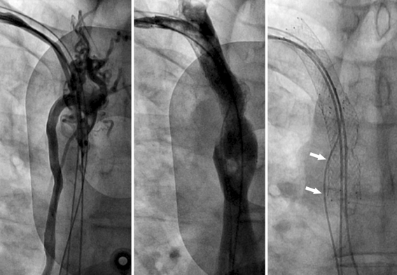
Fig. 5.
Left panel shows angiogram from right subclavian vein, illustrating occlusion of the vena cava superior and collateral circulation through the azygos vein. Middle panel shows the angiographic result after angioplasty and stenting of the vena cava superior. Radiographic appearance of both stents and the position of the newly implanted atrial and ventricular lead are shown in the right panel. The coronary sinus was re-used but functioned properly although the lead was trapped between the stent and the vessel wall (arrows) as illustrated here

