
Figure 1.
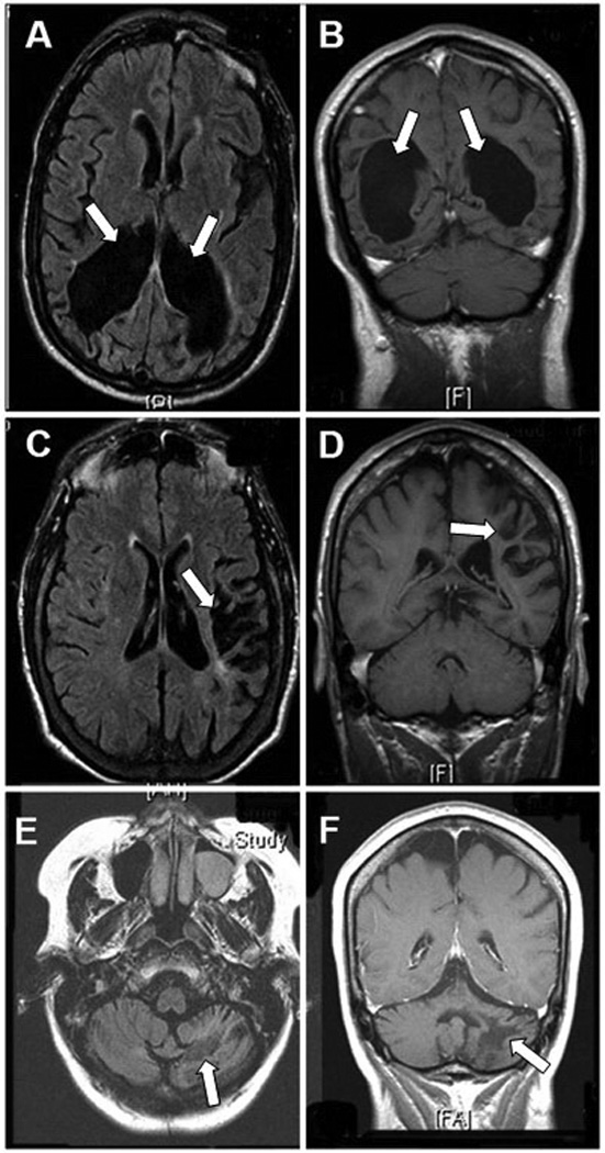
Magnetic resonance imaging of chronic aspects of long survivors of PML: axial FLAIR (A) and coronal T1-weighted images (B) showing marked ventricular enlargement secondary to white matter destruction in the occipital lobes of patient # 10 (arrows) 11 years from onset of PML symptoms with preservation of the occipital gray matter. In patient # 24, there is focal left fronto-parietal subcortical atrophy in axial FLAIR (C) and coronal T1-weigted images (D) (arrows) with preservation of the cortical ribbon after 13 years of evolution. Patient # 6 has extensive destruction of the left cerebellar white matter 10 years from PML diagnosis with atrophy in axial FLAIR (E) and coronal T1-weighted images (F) (arrows). None of these chronic lesions enhanced after administration of gadolinium (not shown).