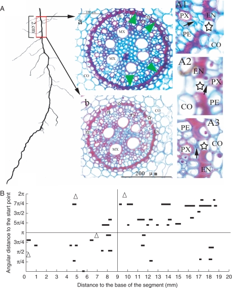
Fig. 6.
Distribution of passage cells in an old root segment near the root base. (a) Cross-sections at about 14 cm (a) and 12 cm (b) from the root tip. Note that the passage cells (indicated by stars) are much less frequent in the section taken near the root base (a) than in the section from the more distal position (b). The green arrowheads indicate the radial position of the four lateral roots on the 2-cm-long segment. (A1), (A2) and (A3) are magnifications of portions of selected sections. Several (A1) or one (A2) small pericycle cells (indicated by black arrows) lie between the protoxylem poles and the passage cells. Some of them are very small and look similar to an intercellular space (A3). CO, Cortex; EN, endodermis; PE, pericycle; MX, metaxylem; PX, protoxylem. (B) The longitudinal arrangement of passage cells along the root axis. Passage cells are arranged in short discontinuous files. They tend to cluster in one half of the transverse plane over a short distance (in 0-π for 0–9 mm from the root base; in π-2π for 9–19 mm from the root base). In the longer distance, passage cells seem to disperse radially. Triangles indicate the positions of the lateral roots.

