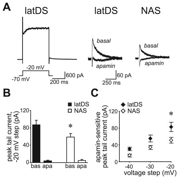
Fig. 4.
Small-conductance calcium-dependent potassium channel function, measured with voltage-clamp, was significantly greater in lateral dorsal striatum (latDS) than in nucleus accumbens shell (NAS) neurons. (A) Depolarizing current steps resulted in a tail current upon return to the −70-mV holding potential. The left trace shows an example from a latDS neuron of the full current response to the −20-mV depolarization. The right traces show examples of the post-depolarization tail current in latDS and NAS neurons, before and after apamin administration. (B) Data for the step to −20 mV showing that the peak of the tail current was significantly greater in latDS than in NAS neurons, and was significantly reduced by apamin in both regions. bas, baseline; apa, apamin. (C) Apamin-sensitive peak tail currents were significantly larger in latDS than in NAS neurons. *P < 0.05.

