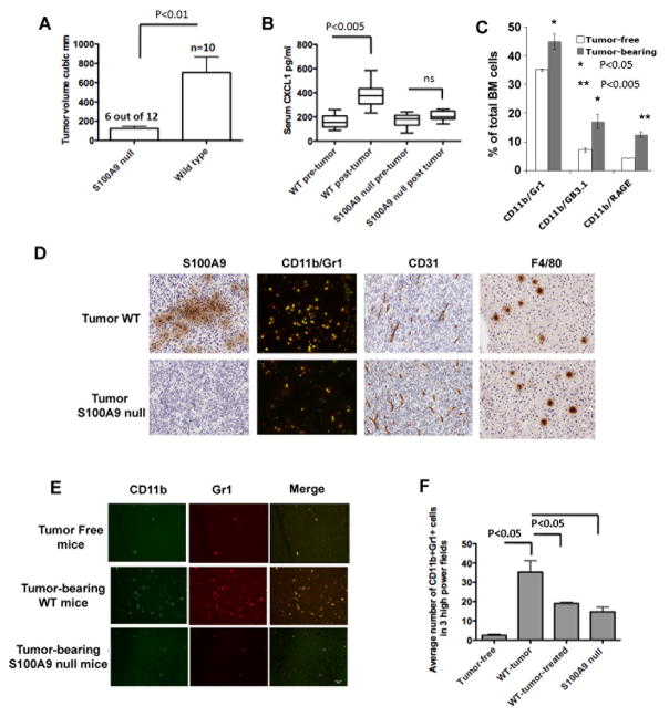
Figure 6.
A. Tumor volumes of ectopic MC38 tumors in wild type (n=10) and S100A9 null mice (n=12) 3 weeks after sc injection of 1×10⁶ cells. 6 out of 12 S100A9 null mice showed significantly reduced tumor growth shown here. In addition, 2 of the remaining six S100A9 null mice completely rejected the tumors. B. CXCL1 in sera of wild type and S100A9 null mice before and 3 weeks after MC38 tumor growth. C. Quantitation of CD11b+ cells co-staining with Gr1 or GB3.1 glycans or RAGE from bone marrow of MC38 tumor-bearing wild type mice D. Tumors were examined for infiltrating macrophages and tumor endothelial cells by immunochemical staining for S100A9+ cells, CD11b+Gr1+ cells (merged images of Alexa-488 stained CD11b+ cells and Alexa-594 stained Gr1+ cells showing double positive yellow cells), and CD31+ and F4/80+ cells (200×). E. Representative sections showing CD11b+Gr1+ cells in premetastatic livers of tumor-free and tumor-bearing mice. F. Quantitation of average number of CD11b+Gr1+ cells in premetastatic livers in 3 high power fields.

