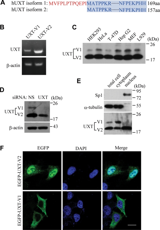
FIGURE 1:
Two isoforms of UXT localize in nucleus and cytoplasm, respectively. (A) Sequence alignment of UXT-V1 against UXT-V2. (B) RT-PCR analysis of UXT mRNA in HeLa cells. (C) Immunoblot of UXT in several cell lines. (D) Immunoblot of UXT in UXT-RNAi HeLa cells or controls. (E) Subcellular fractionation analysis in HeLa cells. Control antibodies indicate accuracy of fractionation (Sp1, nucleus; α-tubulin, cytoplasm). (F) Confocal microscopy analysis of HeLa cells transfected with UXT-V1 or UXT-V2, with EGFP tagged at the N terminus. Scale bar: 10 μm.

