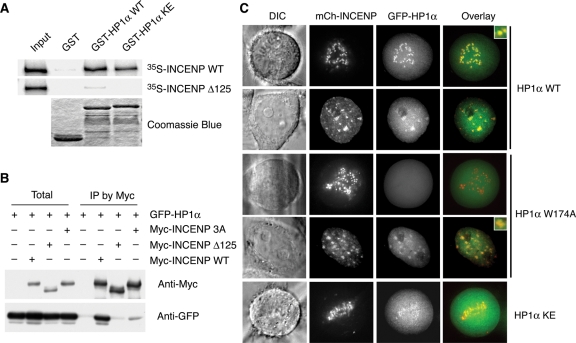
FIGURE 1:
INCENP binding and mitotic centromere localization of HP1 require the CSD. (A) Recombinant purified GST, GST-HP1α WT, or GST-HP1α hinge mutant (KE; K89E, R90E, and K91E) on glutathione-agarose beads were incubated with in vitro translated 35S-labeled Myc-INCENP WT or Δ125. Bound fractions were analyzed by SDS–PAGE followed by autoradiography and Coomassie blue staining. (B) HeLa tet-on cells were transfected with plasmids encoding GFP-HP1α and Myc-INCENP WT, Δ125, or the PXVXL/I motif mutant (3A; P167A, V169A, and I171A). Cell lysates and the α-Myc IP were blotted with α-Myc and α-GFP. (C) HeLa tet-on cells were transfected with plasmids encoding mCherry-INCENP and GFP-HP1α WT, W174A, or KE and monitored with live-cell imaging. mCherry-INCENP and GFP-HP1α signals are shown in red and green, respectively, in the overlay. DIC, differential interference contrast.

