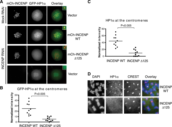
FIGURE 2:
INCENP recruits HP1α to mitotic centromeres. (A) HeLa tet-on cells were first cotransfected with mCherry-INCENP and GFP-HP1α for 6 h and then either mock transfected or transfected with INCENP siRNA for another 48 h. Cells were examined with live-cell imaging. mCherry-INCENP and GFP-HP1α signals are shown in red and green, respectively, in the overlay. (B) Quantification of the mitotic centromeric signals of GFP-HP1α of cells (N = 10 cells) in (A). (C) HeLa tet-on cells that stably express mCherry-INCENP WT or Δ125 were transfected with INCENP siRNA for 48 h. Metaphase chromosome spread was prepared from these cells and stained with DAPI, CREST, and α-HP1α. Staining intensities of HP1α at the centromeres were quantified (N = 10 cells). (D) Representative images of the metaphase chromosome spread described in (C). DAPI, HP1α staining, and CREST staining are colored blue, green, and red, respectively, in the overlay.

