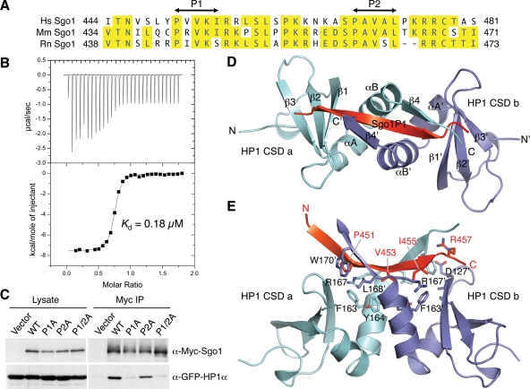
FIGURE 4:
Binding Sgo1 to HP1 involves HP1 CSD and a PXVXL/I motif in Sgo1. (A) Sequence alignment of the HP1-binding region of human (Hs), mouse (Mm), and rat (Rn) Sgo1. The two PXVXL/I motifs are labeled P1 and P2. (B) ITC measurement of the binding between HP1β CSD and Sgo1P1. (C) HeLa tet-on cells were transfected with the GFP-HP1 plasmid together with plasmids encoding Myc-Sgo1 WT, P1A (P451A, V453A, and I455A), P2A (P469A, V471A, and L473A), or P1/2A (P451A, V453A, I455A, P469A, V471A, and L473A) for 24 h and then treated with nocodazole for another 18 h. Cell lysates and Myc IP were blotted with α-Myc and α-GFP. (D) Ribbon drawing of the structure of HP1β CSD–Sgo1P1. Sgo1P1 is colored red, and the two CSD monomers are colored cyan and blue, respectively. (E) Ribbon drawing of the structure of HP1β CSD–Sgo1P1 in an orientation different from (C) and with key binding residues shown in sticks and labeled.

