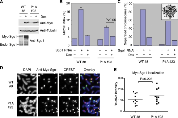
FIGURE 5:
The Sgo1–HP1 interaction is dispensable for Sgo1 localization and sister-chromatid cohesion. (A) HeLa tet-on cells that stably express Myc-Sgo1 WT (clone #8) or P1A (clone #23) under the control of doxycycline were cultured in the absence (–) or presence (+) of doxycycline (Dox) and transfected with Sgo1 siRNA for 24 h. Cell lysates were blotted with α-Myc, α-tubulin, and α-Sgo1. (B) The mitotic index of cells in (A). Cells were stained with propidium iodide and α-H3-pS10 and analyzed by FACS. Ten thousand events were counted for each sample. Mitotic cells have 4N DNA content and are H3-pS10-positive. The average and SD of three experiments are shown. (C) The extent of sister-chromatid separation of cells in (A) as determined by metaphase spread with Giemsa staining (N = 100 cells). A representative image of a cell with separated chromosomes is shown in inset. (D) Metaphase spread of cells in (A) was stained with DAPI (blue in overlay), Myc (green in overlay), and CREST (red in overlay). (E) Quantification of Myc-Sgo1 staining intensities at the centromeres for cells in (D) (N = 10 cells).

