FIGURE 5:
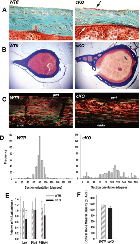
Abnormal cortical bone properties in cKO mice. (A) At 1 mo of age, Goldner trichrome stain show thinner cKO cortices relative to WTfl, but no abnormal osteoid. Also note the prominent osteoblast layer in the periosteum (black arrow) and endosteum (white arrow) in cKO sections. (B) Low-magnification Goldner trichrome-stained cross-sections showing increased cortical porosity and larger cross-sections in cKO bone relative to WTfl. (C) Birefringence of picrosirius red stained cortical sections show more collagen disorganization and a woven bone aspect in cKO relative to WTfl, especially on the periosteal side. (D) Collagen fiber orientation was quantitated by measuring the angle of extinction of polarized light in picrosirius red stained sections. Whereas collagen fiber orientation clusters around 90 degrees in the WTfl, there is a wide, uneven spread of collagen fiber orientation across the whole range (n = 4). (E) Quantitative real-time PCR of femoral bone (without bone marrow) RNA extracts, revealing lower abundance of Lox mRNA, but unchanged Plod and P3HA4 mRNA in cKO relative to WTfl. (F) Cortical bone mineral density (gHA/cc) measured by μCT showing a significant reduction in the cKO relative to WTfl (*, P < 0.05 vs. WTfl, t-test for unpaired samples; n = 4–6).

