FIGURE 1:
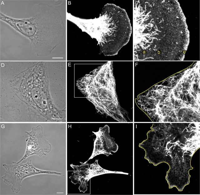
(A–C) Vimentin in a motile 3T3 cell with a typical leading lamellipodium and a trailing tail region. Long VIF are abundant in the tail, in the perinuclear regions, and appear to terminate in the proximal regions of the lamella (C, a). Short filaments (squiggles) are present in the proximal portion of the lamella (C, b), and particles are evident throughout the lamella. The lamellipodium is enriched in vimentin particles (C, c; A, phase contrast; B, C, immunofluorescence using anti-vimentin. C is a higher magnification of B). (D–F) Serum-starved 3T3 cells have mainly rigid-appearing edges devoid of lamellipodia (D). VIF extend to the edge of the cell (D, phase contrast; E, immunofluorescence with anti-vimentin; F is a higher magnification of the region in the box in E; cell edge traced in yellow). (G–I) Within minutes of the addition of serum to serum-starved 3T3 cells, areas of membrane ruffling/lamellipodia become apparent (G). After 30 min, immunofluorescence reveals that the distribution of the different forms of vimentin in the lamella/lamellipodia and tail regions are indistinguishable from those seen in fibroblasts maintained in serum (G, phase contrast; H, anti-vimentin; I, higher magnification of boxed area in H; cell edge traced in yellow). Bars = 10 μm.

