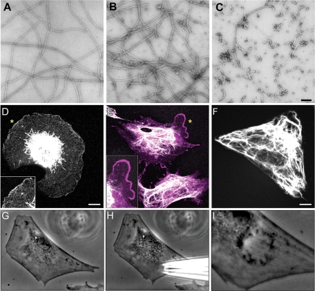
FIGURE 6:
The mimetic 2B2 peptide disassembles VIF in vitro and induces ruffling in vivo. (A–C) The addition of the mimetic 2B2 peptide to recombinant human vimentin assembled for 1 h (A) induced depolymerization that began within 10 s after addition (B) and rapidly proceeded so that after 5 min predominantly ULF-like structures remained (C). Negative stain electron microscopy. Bar = 100 μm. (D–F) In response to the microinjection of higher concentrations of 2B2 into serum-starved (72 h) mEF cells, the VIF frequently retracted from the entire cell periphery (D, immunofluorescence with anti-vimentin; inset, higher magnification of region indicated by asterisk). Note the presence of vimentin particles in the region between the retracted VIF and the cell surface, which shows extensive membrane ruffling. This cell was fixed 60 min after microinjection. (E) In cells injected with lower concentrations of peptide and fixed 30–60 min later, the VIF frequently disassembled near the injection site, where lamellipodia also formed, as indicated by Arp2/3 staining (E, double-label immunofluorescence; vimentin, white; Arp2/3, magenta; inset, higher magnification of region near asterisk). In comparison, control serum-starved (72 h) 3T3 cells or mEF that were microinjected with a scrambled peptide, BSA, or buffer alone did not show an altered distribution of their VIF and lamellipodia were not induced (F, GFP-vimentin in a live 3T3 cell after the injection of scrambled peptide). (G–I) Occasionally, upper surface ruffles were apparent after the microinjection of 2B2 at the injection site (G, H, live 3T3 cell before; I, 10 min postinjection). Bars = 10 μm.

