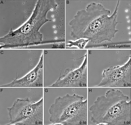
FIGURE 8:
Live imaging of altered motility following VIF disassembly in 2B2-microinjected serum-deprived cells. (A–H, live phase contrast images of the same cell). Moving mEF cells maintained in 2% serum were microinjected with 2B2 opposite ruffling areas (A, arrow marks the microinjection site). Small membrane protrusions became apparent near the site of microinjection within ∼10 min (D, 15 min). This lamellipodium became more prominent with time, and eventually this cell began to move in the direction of the newly formed lamellipodium (A–H, note the retraction fibers above the cell in A and below it in B; see Supplemental Video S4). Bar = 100 μm.

