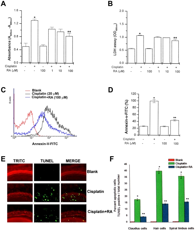
Figure 4. RA inhibits expression of apoptotic marker by cisplatin.
HEI-OC1 cells (1×10⁶/well) were treated with various concentrations of RA (1, 10, and 100 µM) for 2 h and then stimulated with cisplatin (20 µM) for 48 h. Internucleosomal DNA fragmentation was quantitatively determined by assaying for cytoplasmic mononucleosome- and oligonucleosome-associated histone accumulated in membrane-intact cells at the indicated time points (A). LDH levels on supernatant were assayed by cytotoxic assay kit (B). Apoptosis was measured by staining with FITC-labeled annexin V, followed by flow cytometric analysis (C), and the percentage of apoptotic cells in the total cell population is shown (D). The basal turns of the cochlea were stained with TRITC-conjugated phalloidin (red), TUNEL (green), and examined under fluorescent microscope. Results are representative of three independent experiments (E). The percentage of apoptotic cells in different type of cells in the explants is shown (F). Data are representative of three independent experiments. Data are mean ± S.D. of three independent experiments performed in duplicate. *P<0.01, compared with unstimulated cells. **P<0.05 compared with cisplatin alone. * and ** represent significance determined by independent t-test.

