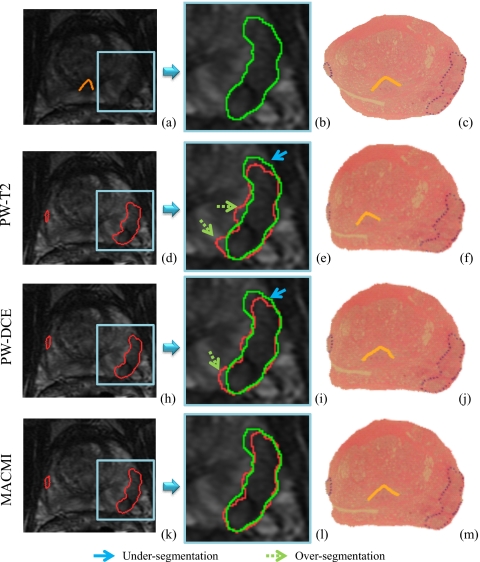
Figure 5.
(a) 3T in vivo T2-w MRI of a prostate with a clearly visible DIL and magnified in (b) with a manual estimate of CaP extent. (c) Closest corresponding WMH slice with CaP ground truth (dotted line) and urethra. [(d) and (e)] T2-w MRI with estimate of CaP extent as mapped from (f) WMH via elastic registration using only T2-w MRI. [(h) and (i)] T2-w MRI with CaP estimate from (j) WMH registered to DCE (T1-w) MRI (coregistered to T2-w MRI). [(k) and (l)] Registration using both T2-w and DCE MRI via MACMI results in closer agreement of the registration-derived CaP extent and the manual estimate. The verumontanum of the urethra is also shown on the registered WMH images in (f), (j), and (m).

