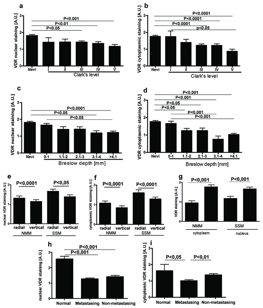
Figure 4.
VDR expression changes during progression of melanocytic lesions and is determined by histological type and aggressiveness of melanomas.
a. VDR nuclear staining in nevi and melanomas assessed according to Clark’s level.
b. VDR cytoplasmic staining in nevi and melanomas assessed according Clark’s level.
c. VDR nuclear staining in nevi and melanomas assessed according Breslow’s thickness.
d. VDR cytoplasmic staining in nevi and melanomas assessed according Breslow’s thickness.
e. VDR immunoreactivity in nuclei of radial and vertical growth phases of nodular (NMM) and superficial spreading (SSM) melanomas.
f. VDR immunoreactivity in cytoplasm of radial and vertical growth phases of nodular (NMM) and superficial spreading (SSM) melanomas.
g. Comparison of VDR expression in nodular (NMM) and superficial spreading (SSM) melanomas.
h. Comparison of VDR expression in nuclei of normal skin and primary metastasing and non-metastasing melanomas.
i. Comparison of VDR expression in cytoplasm of normal skin and primary metastasing and non-metastasing melanomas.
A.U. – arbitrary units.

