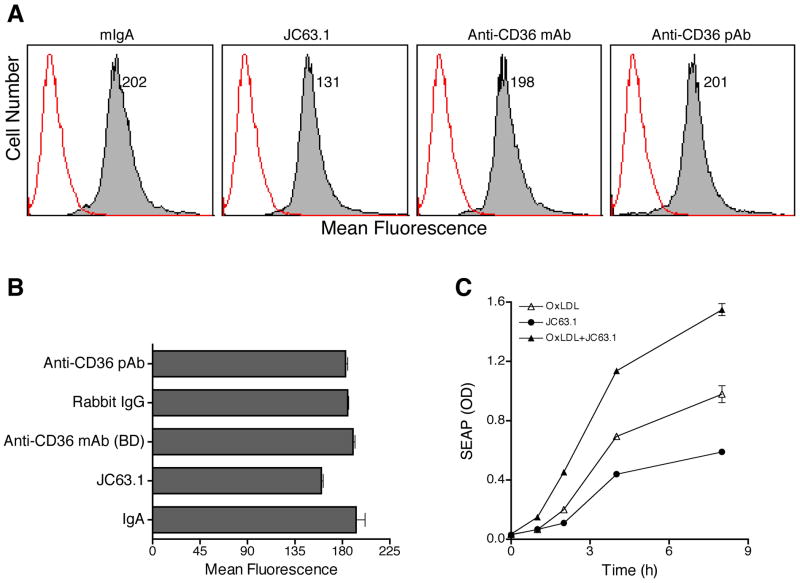
Figure 7. Activating mCD36 mAb enhances oxLDL-induced macrophage activation.
OxLDL binding to RAW-Blue cells were determined by flow cytometry (A and B). Cells were treated with mIgA, activating mCD36 mAb (JC63.1), anti-mCD36 mAb, or anti-mCD36 pAb. OxLDL binding (closed histogram) was detected using goat anti apoB IgG-biotin followed by streptavidin-PE. Cells treated identically without oxLDL (open histogram) were used as a negative control. Number indicates mean fluorescence. C, Activating mCD36 mAb enhances oxLDL-induced macrophage inflammatory response. RAW-Blue cells were co-incubated with activating mCD36 mAb (2 μg/ml) and oxLDL (5 μg/ml). Cells treated with either one of the reagents were used to determine the basal activation. SEAP activity in the supernatant was analyzed to determine kinetics of inflammatory response.

