
Fig. 1.
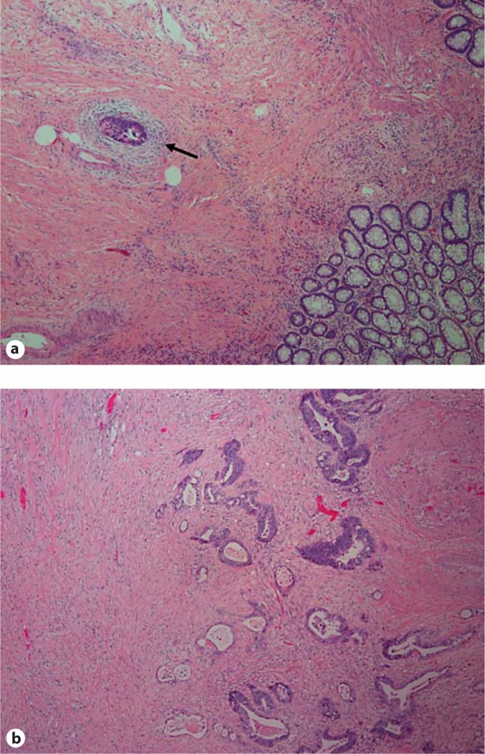
Pathologic response after treatment. Pathologic response was evaluated in all patients. Two representative hematoxylin and eosin-stained sections are shown (40× magnification). a At the time of surgical resection and pathologic evaluation, no gross tumor was identified. Microscopically, there was marked fibrosis with small foci of residual tumor (arrow). This response was classified as RCRG1. b Gross inspection revealed an ulcerated, firm mass consistent with tumor. Microscopically, there was fibrosis with residual tumor. This response was classified as RCRG2.