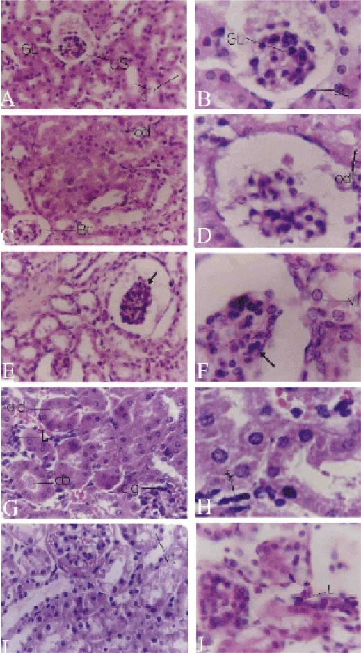
Figure 2.
Light micrographs of kidney. A,B Control kidney with Bowman's capsule with peripheral squamous epithelium(sq), glomerulus (GL), urinary space (US) and normal convoluted tubules (c). C,D T1 group kidney treated with piroxicam for one week with shrinked glomeruli, widened urinary space of the Bowman's capsule and oedema (od). E,F T2 group kidney treated for 2 weeks appearing with shrunken glomeruli (arrow), vacuolated tubules and darkly stained nuclei of the mesangial cells and vacuolations (vt) of the kidney tubules. G,H T3 group kidney treated for 3 weeks appearing with congested glomeruli (cg), odema of kidney tubules (od), inflammation between tubules (L), cell debris inside the tubules (db) and fibroblasts (fr). I,J T4 group treated for 4 weeks appearing with congested glomeruli, vacuolated kidney tubules (vt) and inflammatory cellular infiltration (L). Sections were stained with H&E. Magnifications, X400 (A,C,E,G&I) and X1000 (B,D,F,H&J).

