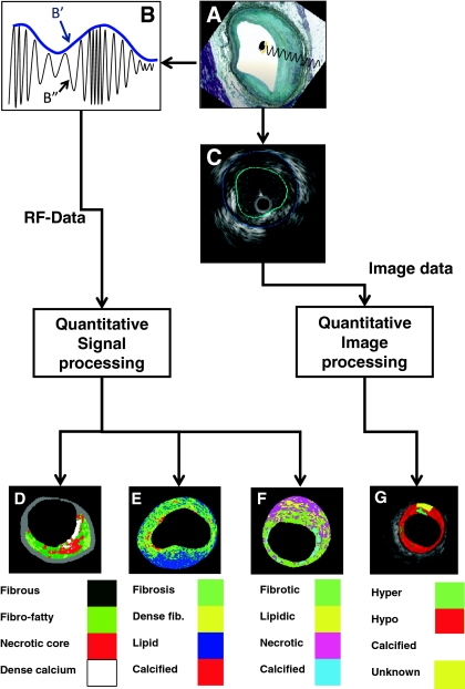
Fig. 1.
A shows how an intravascular ultrasound signal is obtained from the vessel wall within an histology image. The greyscale IVUS image, as can be appreciated in C, is formed by the envelope (amplitude) (B′) of the radiofrequency signal, which is illustrated in B. By greyscale atherosclerotic plaque can be classified into 3 categories: hypoechogenic, hyperechogenic and calcified. A 4th category is defined as unknown, which is tissue that is acoustically showed (G). C shows a cross-sectional view of a grayscale image. The blue lines limit the actual atheroma. The frequency and power of the signal commonly differ between tissues, regardless of similarities in the amplitude. From the backscatter radiofrequency data (identified by B″ in B) different types of tissue information can be retrieved: virtual histology (D), integrated backscattered (IB) IVUS (E) and iMAP (F). Virtual histology is able to detect four tissue types: necrotic core, fibrous, fibro-fatty and dense calcium. The tissues characterized by integrated backscattered (IB) IVUS are lipidic, fibrous and calcified; and iMAP detects fibrotic, lipidic, necrotic and calcified

