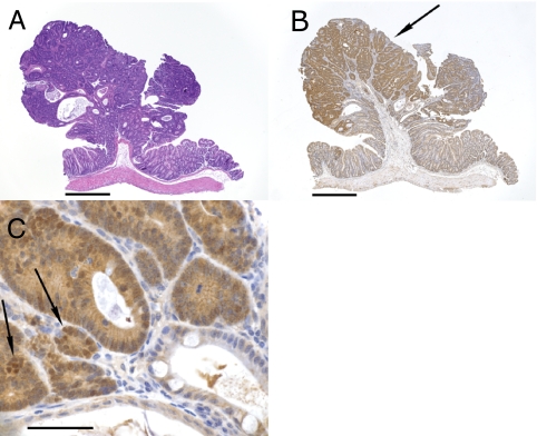
Fig. 1.
A pedunculated adenoma stained with H&E (A) or immunostained for β-catenin (B and C). (B) There is increased staining for β-catenin (arrow) in the adenoma. (C) Higher-power magnification of a different section showing increased cytoplasmic and nuclear (arrows) staining for β-catenin in tumor cells compared with adjacent normal tissue seen in lower right and bottom of picture. (Scale bars: A and B, 500 μm; C, 50 μm.)

