Fig. 1.
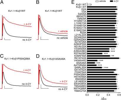
Residues 33–50 of the Kvβ1 N terminus mediate redox modulation. (A–D) K+ current recorded on inside-out patches expressing Kv1.1 with (A, B) Kvβ1 WT, (C) Kvβ1 P55A-Q56A, or (D) Kvβ1 I45A-I46A. In each case, the black trace was recorded before 4-CY or vehicle (1% ethanol) perfusion. After perfusion, a patch was held at −100 mV and its current level was monitored every 30 s with a 200-ms pulse to +60 mV until the current level reached steady state. The solution was then exchanged to the normal inside buffer and the red trace was recorded. This protocol was used for current traces shown in all figures. Scale bars represent 300 pA and 10 ms. Only the first 40 ms of each trace is shown, and the full 200-ms traces are shown in Fig. S1. (E) Bar graph of the 4-CY effect (ΔIss) on alanine mutations to residues 2–70 of the Kvβ1 N terminus. The error bars are SEM from 5 to 15 independent patches. **P < 0.01, ***P < 0.001 vs. Kvβ1 WT by 4-CY; unpaired Student's t test.

