Fig. 3.
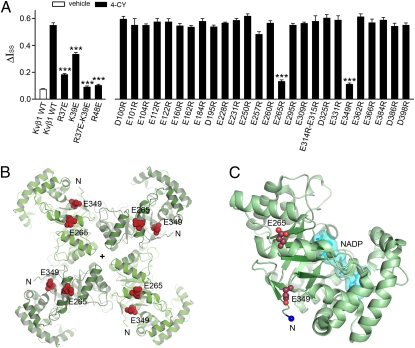
Charged residues on the RRS and Kvβ1 core mediate redox modulation. (A) The 4-CY effect (ΔIss) measured on charge reversal mutations on the RRS and the core domain of Kvβ1. The error bars are SEM from 3 to 15 independent patches. ***P < 0.001 vs. Kvβ1 WT by 4-CY; unpaired Student's t test. (B) The tetrameric core domain of Kvβ2 as viewed looking down the fourfold axis, which is marked with a “+” symbol, from the membrane-facing side. The two glutamate residues, equivalent to E265 and E349 in Kvβ1, are shown as red space-fill. The N terminus of the Kvβ2 core is labeled with an N for each subunit. (C) A monomer of Kvβ2 core, with NADP shown in cyan and the N terminus of the Kvβ2 core marked with a blue sphere. Representative currents for three mutations are shown in Fig. S4.

