
Figure 2.
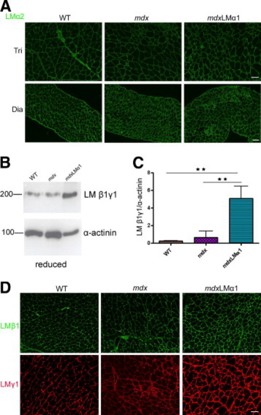
Expression of other laminin chains in mdxLMα1 and mdx mice. A: Immunostaining with laminin α2 chain antibody reveals no reduction in laminin α2 chain expression in mdx and mdxLMα1 muscles, compared with the wild type. B and C: Immunoblotting of skeletal muscle tissue extracts from wild-type (n = 3), mdx (n = 3), and mdxLMα1 mice (n = 3) reveals significant 5-fold upregulation of laminin β1 and γ1 subunits (both at approximately 200 kDa) in mdxLMα1 muscles, compared with both wild-type muscle (**P < 0001) and mdx muscle (**P < 0001). One-way analysis of variance followed by Bonferroni's multiple comparison test was used for statistical analysis. Results are reported as means ± SD. D: Increased immunofluorescent signals for laminin β1 and γ1 chains in mdxLMα1 muscle basement membranes, compared with wild-type and mdx basement membranes. They form heterotrimers with α subunits. Scale bars: 50 μm (A, all images in the same row, and D, all images).