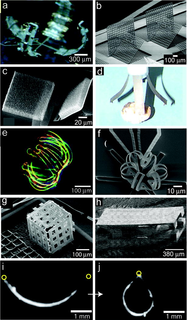
Figure 5. Implementation of various self-folding techniques.

a) Pneumatically actuated microhand. b) SEM micrograph of magnetically assembled microstructures with elastic hinges, where the vertical plates are held in place by the angled lock-in plates. c) SEM micrograph of two TiN membranes with a carpet of Ni-tipped CNTs on top of the membrane partially-folded due to stresses in the TiN hinge. When an external magnetic field is applied, the membrane fully folds 180°. d) Photograph of an end effector gripper using four 0.1 g fingers made of ionic polymer-metal composites. e) Optical micrograph of a 3D microwell with multiple extensions self-folded from a hydrogel bilayer. f) SEM micrograph of an SU8/DLC normally-closed microcage, which folds initially due to residual stress and then opens when thermally actuated. g) SEM micrograph of a microcube fabricated using thermal shrinkage of polyimide. h) SEM micrograph of an SMA actuated microgripper. i) Side view of a gripper composed of muscular thin films with lengthwise-aligned anisotropic myocardium (on the concave surface) that draw the tips together upon contraction j). The circles indicate the ends of the gripper.
a) Reproduced with permission from Reference [101]. Copyright 2006, American Institute of Physics. b) Reproduced with permission from Reference [102]. Copyright 2005, IEEE. c) Reproduced with permission from Reference [108]. Copyright 2008, American Institute of Physics. d) Reproduced with permission from Reference [112]. Copyright 1998, IOP Publishing. e) Reproduced with permission from Reference [111]. Copyright 2005, American Chemical Society. f) Reproduced with permission from Reference [122]. Copyright 2006, Elsevier. g) Reproduced with permission from Reference [123]. Copyright 2007, Springer. h) Figure courtesy of Lawrence Livermore National Laboratory (LLNL), Reference [189]. i,j) Reproduced with permission from Reference [135]. Copyright 2007, AAAS.
