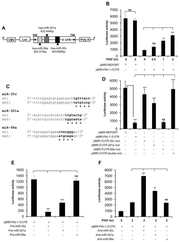
Figure 2. PlGF induced PAI-1-3′UTR reporter activity is abrogated by miR-30c and miR-301a, but not by miR-99a.
(A) Schematic representation of PAI-1-3′UTR luciferase reporter plasmid. The region between nucleotides 292 to 746 of PAI-1-3′UTR containing predicted target binding sites for miR-30c, miR-301a and miR-99a was cloned at the 3′end of luciferase reporter gene in pMIR-REPORT plasmid. (B) HPMVEC were transfected with either pMIR-REPORT control plasmid or pMIR-PAI-1-3′UTR construct. Following transfection, the cells were treated with PlGF for indicated time periods. (C) The sequences of predicted miR-30c, miR-301a and miR-99a binding sites within PAI-1-3′UTR. The bases in bold highlight “seed sequences” and the point mutations in seed sequence are indicated by asterisks. (D) HPMVEC were transfected with either pMIR-REPORT control plasmid, wild-type pMIR-PAI-1-3′UTR or indicated PAI-1-3′UTR mutant constructs for the binding of either miR-30c or miR-301a or miR-99a or both (miR-30c and miR-301a). 24 hr post-transfection, cells were lysed in reporter lysis buffer as per manufacturer’s instructions for luciferase assay. (E) HPMVEC were transfected with pMIR-PAI-1-3′UTR plasmid and indicated pre-miRNA precursor oligonucleotide. (F) HPMVEC were transfected with pMIR-PAI-1-3′UTR plasmid and indicated anti-miR followed by PlGF treatment for 2 hr. (B, D, E and F) Each construct was used at 0.5 μg. All luciferase reporters were co-transfected with β-galactosidase plasmid for normalization of transfection efficiency. The luciferase and_β-galactosidase activities were estimated as described in Materials and Methods. Data are representative of three independent experiments. *, p< 0.05, **, p< 0.01, ***, p< 0.001. ns, not significant.

