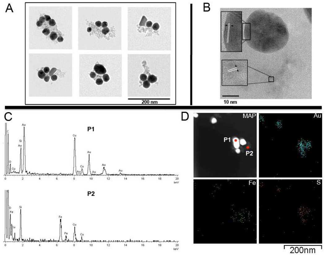
Figure 2. Electron microscopy images of AuMN-DTTC using LWTEM and HRTEM/STEM/EDS.
A) Transmission electron microscopy images of AuMN-DTTC suggests that the complex consists of electron-dense AuNP associated with less electron-dense dextran-coated MN. There are several gold nanostructures per probe B) High resolution-TEM (HRTEM) images show that the metallic lattices of AuNPs and dextran coated MN are located close to each other. The arrows in the magnified insets show two different metallic lattices C) EDS spectra of the dark and light colored region in the EDS map show that the nanostructures are composed of gold and iron. D) Elemental map of the probe. The gold and sulfur components co-localize with the dark colored region. The iron element co-localizes with the light-colored region of the map and is closely associated with the gold.

