
FIGURE 2.
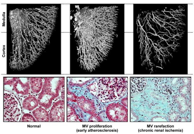
Micro-CT imaging of vascular casts obtained from a swine model of atherosclerotic renal artery stenosis. Atherosclerosis produced by cholesterol feeding induces small vessel proliferation and disturbed endothelial function (middle panel). The kidney beyond a main renal arterial occlusive lesion induced by copper stent experiences dropout (“rarefication”) of small vessels within both cortex and medulla and accelerated tissue fibrosis. From Lerman and Chade, with permission 90.