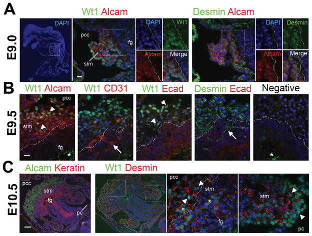
Fig. 1.
Expression of Alcam, desmin, and Wt1 in the STM of the mouse embryos from E9.0 to E10.5. Serial sections prepared from embryos at E9.0 (A, sagittal sections), E9.5 (B, transverse sections), and E10.5 (C, transverse sections) were immunostained with antibodies against Alcam, CD31, desmin, E-cadherin (Ecad), cytokeratin (Keratin), and Wt1. Nuclei were counterstained with DAPI. Note that the STM expresses Alcam, desmin, and Wt1 from E9.0 to E10.5. The nuclear staining of Wt1 is seen in the STM (arrowheads), but not in CD31+ endothelial cells and E-cadherin+ endoderm in E9.5. The nuclear Wt1 expression becomes weak in the STM near the foregut endoderm (fg). Arrows indicate desmin+ Wt1− mesenchymal cells and CD31+ Wt1− endothelial cells trapped in the growing endoderm. Asterisks indicate non-specific signals at the yolk and blood cells. Negative control without primary antibodies is shown in B (negative). pc, peritoneal cavity; pcc, pericardial cavity; stm, septum transversum mesenchyme. Bar, 10 μm (A,B), 50 μm (C).

