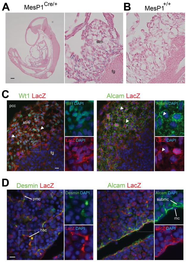
Fig. 3.
MesP1+ mesoderm gives rise to HSCs via STM during mouse embryogenesis. The embryos from the MesP1Cre and Rosa26lacZflox mice were analyzed by X-gal staining (A,B) and immunohistochemistry (C,D). (A) The E9.0 MesP1Cre/+; Rosa26lacZflox/+ embryo shows lacZ expression in the STM adjacent to the foregut endoderm (fg). (B) No lacZ staining in the wild type littermate. Embryos were counterstained with eosin. (C) The STM in E9.5 MesP1Cre/+; Rosa26lacZflox/+ embryos coexpresses lacZ with Wt1 or Alcam (arrowheads). (D) Desmin+ HSCs and PMCs and Alcam+ MCs and SubMCs coexpresses lacZ in E12.5 MesP1Cre/+; Rosa26lacZflox/+ embryos. Nuclei were counterstained with DAPI. hsc, hepatic stellate cells; mc, mesothelial cells; pcc, pericardial cavity; pmc, perivascular mesenchymal cells; stm, septum transversum mesenchyme; submc, submesothelial cells. Bar, 100 μm (A), 10 μm (C,D).

