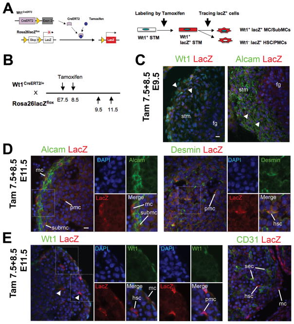
Fig. 4.
The Wt1+ STM gives rise to HSCs and PMCs during liver development. The Wt1+ STM lineage was analyzed using the Wt1CreERT2 and Rosa26lacZflox mice. (A) Tamoxifen induces lacZ expression in Wt1+ STM. After tamoxifen injection, the CreERT2 excises the stop sequence between the loxP sites (triangles). Then, the lacZ gene is expressed in Wt1-expressing cells. If the STM gives rise to HSCs and PMCs, tamoxifen injection results in the expression of lacZ in Wt1− HSCs and PMCs inside the liver. (B) After tamoxifen injection twice at E7.5 and 8.5, the embryos at E9.5, and 11.5 were analyzed. (C-E) Immunohistochemistry of Alcam, CD31, desmin, lacZ, and Wt1 in the E9.5 (C) and E11.5 (D,E) embryos. Nuclei were counterstained with DAPI. Arrowheads indicate lacZ+ cells in Wt1+ or Alcam+ cells in the STM (C). Note that lacZ signals are detected in 5.6 ± 1.0% of Alcam+ cells in the STM in the E9.5 embryos. The percentage was obtained from total 906 Alcam+ cells. Results are means ± SD of 5 independent sections. In E11.5 embryos, lacZ expression is seen in Alcam+ MCs and SubMCs (D). LacZ is expressed in 10.5 ± 4.9% (ML) and 9.0 ± 2.7% (LL) of desmin+ cells in the E11.5 livers. These percentages were obtained from 1,643 (ML) and 2,058 (LL) desmin+ HSCs and PMCs inside the liver. No Wt1 expression in the lacZ+ HSCs and PMCs (E, arrowheads). No lacZ expression in CD31+ SECs (E). fg, foregut endoderm; hsc, hepatic stellate cells; mc, mesothelial cells; pmc, perivascular mesenchymal cells; sec, sinusoidal endothelial cells; stm, septum transversum mesenchyme; submc, submesothelial cells. Bar, 10 μm.

