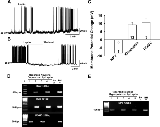
Fig. 3.
Leptin excites kisspeptin and POMC neurons but inhibits NPY neurons. A, Leptin (100 nm) depolarized a kisspeptin neuron and increased the firing frequency, which continued even after a long “washout” period for 30 min until the cell was harvested for scRT-PCR. Leptin had a similar long-lasting depolarizing effect on POMC neurons (data not shown). B, In contrast, leptin (100 nm) hyperpolarized NPY neurons (five of five) and abrogated firing, which fully recovered after several minutes of washing out leptin. C, Summary of the effects of leptin on NPY, kisspeptin, and POMC neurons. Leptin depolarized kisspeptin and POMC neurons, 9.8 ± 1.4 mV and 10.8 ± 2.0 mV, respectively. In contrast, leptin hyperpolarized both male (n = 2) and female (n = 3) NPY neurons (8.3 ± 1.8 mV, n = 5). D and E, Representative gels illustrating the expression of kisspeptin (Kiss-1), Dyn, and POMC mRNAs in leptin-depolarized neurons (D) or NPY mRNA expression in leptin-hyperpolarized neurons (E). Basal hypothalamic total RNA was also reverse transcribed in the presence or absence of RT (tissue controls, +/− respectively). Two different ladders (L) were used: one for Kiss1 (50-bp DNA ladder) and the other for Dyn, POMC, and NPY (100-bp DNA ladder).

